Message Board

Respected readers, authors and reviewers, you can add comments to this page on any questions about the contribution, review,        editing and publication of this journal. We will give you an answer as soon as possible. Thank you for your support!

Name
E-mail
Phone
Title
Content
Verification Code
Volume 38 Issue 3
May  2020
Turn off MathJax
Article Contents

SHANG Liuwenxin, SUN Xin, PENG Cheng, XIE Hehui, CHEN Fengyuan, ZHANG Chuan. Improvement of impaired endothelial progenitor cell functions by in vitro drug interference in DOCA-salt hypertensive mice[J]. Journal of Pharmaceutical Practice and Service, 2020, 38(3): 221-226. doi: 10.12206/j.issn.1006-0111.201912074
Citation: SHANG Liuwenxin, SUN Xin, PENG Cheng, XIE Hehui, CHEN Fengyuan, ZHANG Chuan. Improvement of impaired endothelial progenitor cell functions by in vitro drug interference in DOCA-salt hypertensive mice[J]. Journal of Pharmaceutical Practice and Service, 2020, 38(3): 221-226. doi: 10.12206/j.issn.1006-0111.201912074

Improvement of impaired endothelial progenitor cell functions by in vitro drug interference in DOCA-salt hypertensive mice

doi: 10.12206/j.issn.1006-0111.201912074
  • Received Date: 2019-12-16
  • Rev Recd Date: 2020-03-05
  • Available Online: 2020-05-20
  • Publish Date: 2020-05-01
  •   Objective  To investigate the effect of tetrahydrobiopterin (BH4), superoxide dismutase-polyethylene glycol (PEG-SOD) and N(G)-nitro-L-arginine (L-NNA) on impaired endothelial progenitor cell (EPC) functions in DOCA-salt hypertensive mice.  Methods  DOCA-salt hypertension was created and systolic blood pressure was measured by tail-cuff methods. EPC was isolated from bone marrow of mice and characterized by flow cytometry and fluorescence microscopy. EPC of DOCA-salt mice was incubated with BH4, PEG-SOD, and L-NNA for 24 h, then in vitro EPC function assays were performed.  Results  Compared with control group, systolic blood pressure was significantly increased in DOCA-salt mice. Both EPC adhesion and angiogenesis functions were impaired in DOCA-salt mice compared to control animals, which were reversed by incubation with BH4, PEG-SOD and L-NNA.  Conclusion  BH4, PEG-SOD and L-NNA rescued the impairments from EPC functions in DOCA-salt hypertensive mice.
  • [1] SIMIONESCU M. Implications of early structural-functional changes in the endothelium for vascular disease[J]. Arterioscler Thromb Vasc Biol,2007,27(2):266-274. doi:  10.1161/01.ATV.0000253884.13901.e4
    [2] SHANTSILA E, WATSON T, LIP G Y. Endothelial progenitor cells in cardiovascular disorders[J]. J Am Coll Cardiol,2007,49(7):741-752. doi:  10.1016/j.jacc.2006.09.050
    [3] KAWAMOTO A, LOSORDO D W. Endothelial progenitor cells for cardiovascular regeneration[J]. Trends Cardiovasc Med,2008,18(1):33-37. doi:  10.1016/j.tcm.2007.11.004
    [4] CHEN D D, DONG Y G, YUAN H, et al. Endothelin 1 activation of endothelin A receptor/NADPH oxidase pathway and diminished antioxidants critically contribute to endothelial progenitor cell reduction and dysfunction in salt-sensitive hypertension[J]. Hypertension,2012,59(5):1037-1043. doi:  10.1161/HYPERTENSIONAHA.111.183368
    [5] XIE H H, ZHOU S, CHEN D D, et al. GTP cyclohydrolase I/BH4 pathway protects EPCs via suppressing oxidative stress and thrombospondin-1 in salt-sensitive hypertension[J]. Hypertension,2010,56(6):1137-1144. doi:  10.1161/HYPERTENSIONAHA.110.160622
    [6] DU Y H, GUAN Y Y, ALP N J, et al. Endothelium-specific GTP cyclohydrolase I overexpression attenuates blood pressure progression in salt-sensitive low-renin hypertension[J]. Circulation,2008,117(8):1045-1054. doi:  10.1161/CIRCULATIONAHA.107.748236
    [7] MARROTTE E J, CHEN D D, HAKIM J S, et al. Manganese superoxide dismutase expression in endothelial progenitor cells accelerates wound healing in diabetic mice[J]. J Clin Invest,2010,120(12):4207-4219. doi:  10.1172/JCI36858
    [8] HAGIWARA H, HIGASHIBATA A, OGAWA S, et al. Effectiveness of endothelial progenitor cell culture under microgravity for improved angiogenic potential[J]. Sci Rep,2018,8(1):14239. doi:  10.1038/s41598-018-32073-2
    [9] 杨栋, 辛世杰, 李凤贺, 等. 小鼠骨髓源性内皮祖细胞的培养和表面标志物鉴定[J]. 中国医科大学学报, 2011, 40(12):1063-1066.
    [10] 杨兆华. 内皮祖细胞(EPCs)对移植性动脉硬化的作用及其机制研究[D]. 上海: 复旦大学, 2009.
    [11] MOENS A L, KASS D A. Tetrahydrobiopterin and cardiovascular disease[J]. Arterioscler Thromb Vasc Biol,2006,26(11):2439-2444. doi:  10.1161/01.ATV.0000243924.00970.cb
    [12] ZHENG J S, YANG X Q, LOOKINGLAND K J, et al. Gene transfer of human guanosine 5′-triphosphate cyclohydrolase I restores vascular tetrahydrobiopterin level and endothelial function in low renin hypertension[J]. Circulation,2003,108(10):1238-1245. doi:  10.1161/01.CIR.0000089082.40285.C3
  • 加载中
通讯作者: 陈斌, [email protected]
  • 1. 

    沈阳化工大学材料科学与工程学院 沈阳 110142

  1. 本站搜索
  2. 百度学术搜索
  3. 万方数据库搜索
  4. CNKI搜索

Figures(4)

Article Metrics

Article views(6010) PDF downloads(21) Cited by()

Related
Proportional views

Improvement of impaired endothelial progenitor cell functions by in vitro drug interference in DOCA-salt hypertensive mice

doi: 10.12206/j.issn.1006-0111.201912074

Abstract:   Objective  To investigate the effect of tetrahydrobiopterin (BH4), superoxide dismutase-polyethylene glycol (PEG-SOD) and N(G)-nitro-L-arginine (L-NNA) on impaired endothelial progenitor cell (EPC) functions in DOCA-salt hypertensive mice.  Methods  DOCA-salt hypertension was created and systolic blood pressure was measured by tail-cuff methods. EPC was isolated from bone marrow of mice and characterized by flow cytometry and fluorescence microscopy. EPC of DOCA-salt mice was incubated with BH4, PEG-SOD, and L-NNA for 24 h, then in vitro EPC function assays were performed.  Results  Compared with control group, systolic blood pressure was significantly increased in DOCA-salt mice. Both EPC adhesion and angiogenesis functions were impaired in DOCA-salt mice compared to control animals, which were reversed by incubation with BH4, PEG-SOD and L-NNA.  Conclusion  BH4, PEG-SOD and L-NNA rescued the impairments from EPC functions in DOCA-salt hypertensive mice.

SHANG Liuwenxin, SUN Xin, PENG Cheng, XIE Hehui, CHEN Fengyuan, ZHANG Chuan. Improvement of impaired endothelial progenitor cell functions by in vitro drug interference in DOCA-salt hypertensive mice[J]. Journal of Pharmaceutical Practice and Service, 2020, 38(3): 221-226. doi: 10.12206/j.issn.1006-0111.201912074
Citation: SHANG Liuwenxin, SUN Xin, PENG Cheng, XIE Hehui, CHEN Fengyuan, ZHANG Chuan. Improvement of impaired endothelial progenitor cell functions by in vitro drug interference in DOCA-salt hypertensive mice[J]. Journal of Pharmaceutical Practice and Service, 2020, 38(3): 221-226. doi: 10.12206/j.issn.1006-0111.201912074
  • 保护血管内皮对维护心血管系统健康有着重要意义[1]。近年的研究证实,血管内皮完整性的维持不仅依赖于现有的内皮细胞,而且与骨髓来源的具有向成熟内皮细胞分化能力的未成熟细胞—内皮祖细胞(endothelial progenitor cell, EPC)的募集有关[2-3]。临床研究证实,外周血中循环EPC的数量与冠心病常见的危险因素(如高血压、糖尿病等)呈负相关关系,EPC可作为治疗缺血、肺动脉高压等血管疾病状态的新靶点[2-3]。本课题组和他人的研究均证实[2, 4-5],高血压动物和患者的EPC均出现显著的功能受损。因此,如果能够通过药物干预手段逆转其受损的EPC功能,无疑对减轻高血压患者与内皮功能相关的靶器官损伤,以及改善高血压EPC移植治疗效果等具有重要意义。

    去氧皮质酮/盐(Deoxycorticosterone acetate-salt, DOCA-salt)高血压模型是一种盐敏感性高血压动物模型,盐敏感性高血压在美国高血压患者中占1/3的比例,是一种重要的高血压类型[4-6]。课题组既往的研究证实,DOCA-salt高血压小鼠EPC功能受损与其内皮型一氧化氮合酶(endothelial nitric oxide synthase, eNOS)的脱偶联(uncoupling)有关[5]。在研究中我们也找到了一些对eNOS脱偶联相关的信号通路/分子有一定改善作用的药物,如四氢生物蝶呤(tetrahydrobiopterin, BH4),超氧化物歧化酶-聚乙二醇(superoxide dismutase-polyethylene glycol, PEG-SOD),N(G)-硝基-L-精氨酸(N(G)-nitro-L-arginine, L-NNA)等[5-6]。但目前尚不知道这些药物是否可有效逆转DOCA-salt小鼠受损的EPC功能。本研究拟在以往研究的基础上,寻找改善盐敏感性高血压小鼠EPC功能失常的有效药物干预手段,为进一步的EPC移植治疗研究奠定基础。

    • 野生型(C57BL/6)雄性小鼠,10~12周龄,体重20~25 g(上海斯莱克实验动物中心)。

    • BH4、PEG-SOD、L-NNA(美国Sigma公司),EGM-2培养基(Lonza公司),胎牛血清(美国GIBCO公司),Matrigel胶(美国BD公司)。

    • 选用10~12周龄,体重20~25 g野生型(C57BL/6)雄性小鼠进行模型制作。小鼠麻醉,侧卧位固定,腹部纵行切口,暴露左肾,小心结扎肾动脉和肾静脉后,进行左肾切除,缝合切口。然后在小鼠两前臂中间的位置开一个大约1 cm的纵行切口,皮下埋置150 mg/kg的DOCA缓释片。DOCA组小鼠给予含1.0% NaCl和0.2% KCl的盐水。假手术组小鼠也进行左肾切除,但不给DOCA缓释片,而且饮用常规自来水。手术后,所有动物被单独饲养于一个干净的塑料笼内。术后第21天用尾动脉测压法测定动物的收缩压水平。

    • 将玻璃粘连蛋白(vitronectin)加入6孔板预处理后备用。小鼠麻醉后处死。使用无菌器械剥离小鼠双侧后肢股骨、胫骨并剔除肌肉组织,于超净台内用EGM-2培养基冲出骨髓。将骨髓细胞浓度调定至2.5×10⁵ /cm2后,加入预处理的6孔板内培养(37 ℃、5% CO2)。第4天弃去原培养基,更换新鲜培养基,第7天使用。

    • 本研究采用了以下两种鉴定方法。

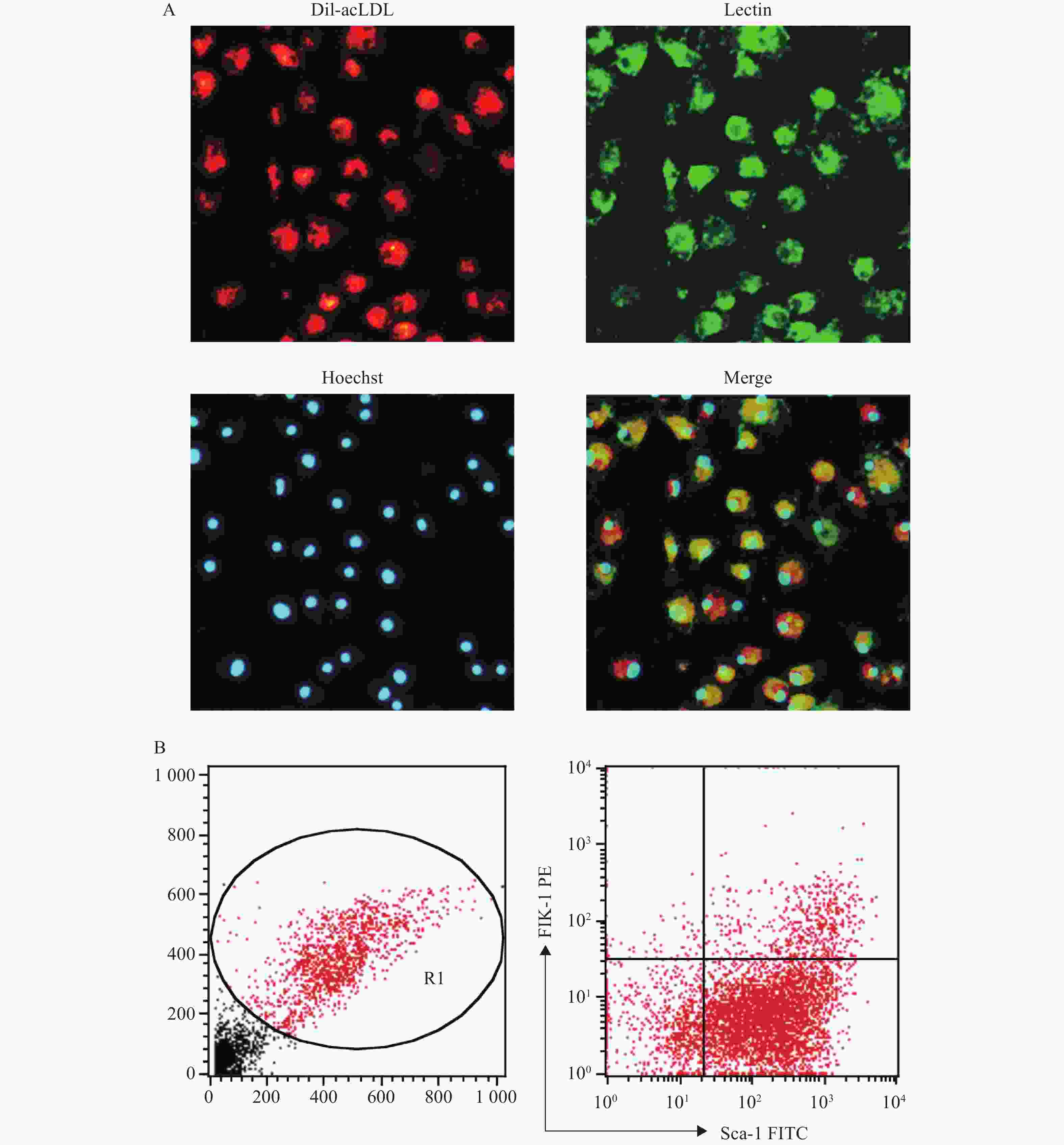
      (1)流式细胞仪法:EPC培养至第7天后,用0.125%胰酶-0.02% EDTA消化,洗涤后调整细胞密度为2×10⁶ /ml,将细胞与Sca-1的抗体(1 µg/ml)和Flk-1的抗体(1 µg/ml)在冰上共孵育1 h,注意避光。之后用5%BSA/PBS洗涤,再用2%的多聚甲醛固定10 min,用流式细胞仪检测Sca-1、Flk-1双标阳性的细胞,即为EPC。

      (2)荧光显微镜法:EPC培养至第7天后,用PBS洗细胞,加入1 mg/ml Dil-acLDL(1:200),于37 ℃避光孵育4 h,用PBS洗细胞,加2%多聚甲醛固定10 min。加10 µg/ml的Lectin,室温避光孵育1 h,再用PBS洗细胞。加Hoechst 33258(10 µg/ml),室温避光孵育30 min,用PBS洗细胞,加2%多聚甲醛固定10 min后,在荧光显微镜下观察Dil-acLDL(红色荧光)、Lectin(绿色荧光)、Hoechst 33258(蓝色荧光)3种染料染色阳性的细胞,即为EPC。

    • 小鼠骨髓来源EPC培养至第6天后换液,分别用BH4 (10 µmol/L), PEG-SOD (100 U/L)和L-NNA (0.8 mmol/L)3种药物孵育24 h后,测定其小管形成功能和黏附功能。

    • 培养7 d的EPC用0.125%胰酶−0.02% EDTA消化,将1×10⁴细胞接种于vitronectin包被好的96孔板中,每个样本设4复孔。37 ℃、5% CO2孵育24 h后,去除培养基,PBS洗涤3次,然后用Hoechst 33258 (5 µg/ml)染细胞核,再用PBS洗涤3次。在荧光显微镜下,随机选取3个视野(100×)计算黏附细胞数并取平均值。

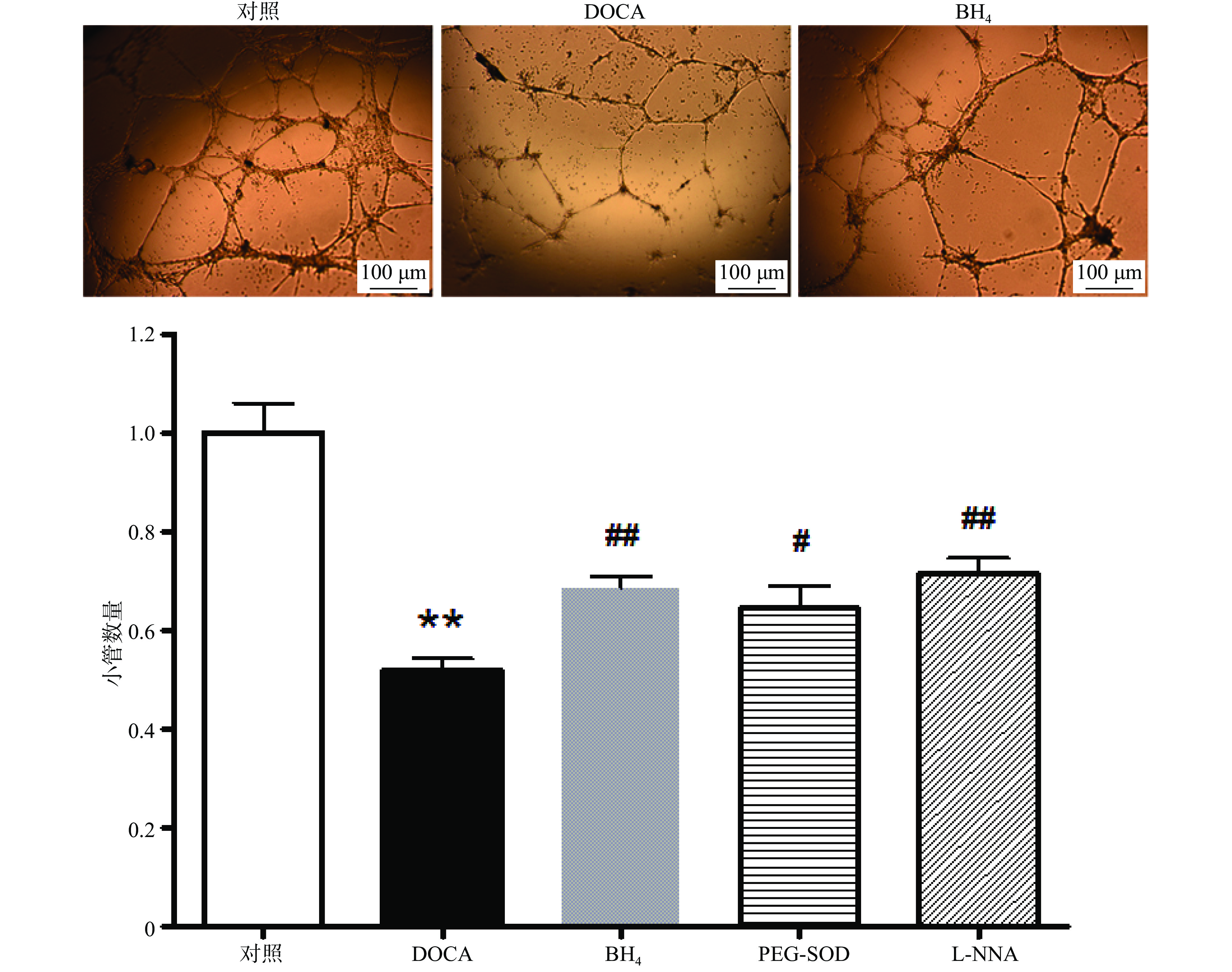
    • 在冰上预冷的96孔板中加入60 µl的matrigel,于37 ℃孵育30 min。培养7 d的EPC用0.125%胰酶−0.02% EDTA消化,将5×10⁴细胞接种于matrigel上,在37 ℃,5% CO2孵箱中孵育6 h后,于显微镜下随机选取4个视野(40×)计算形成小管的数目并取平均值。

    • 所得实验数据均通过Prism统计软件进行分析,分别选取配对t-检验和单因素方差分析(One-Way ANOVA)作为2组数据和3组以上数据之间比较的统计学分析方法;选用Newman Keuls法进行均数之间的多重比较。数据书写格式为(均数±标准误)。以P<0.05表示差异有统计学意义。

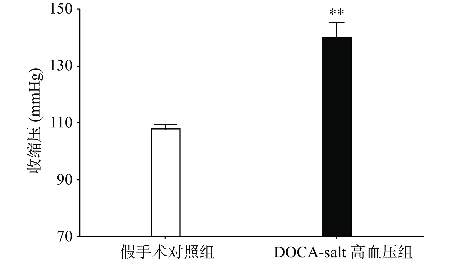
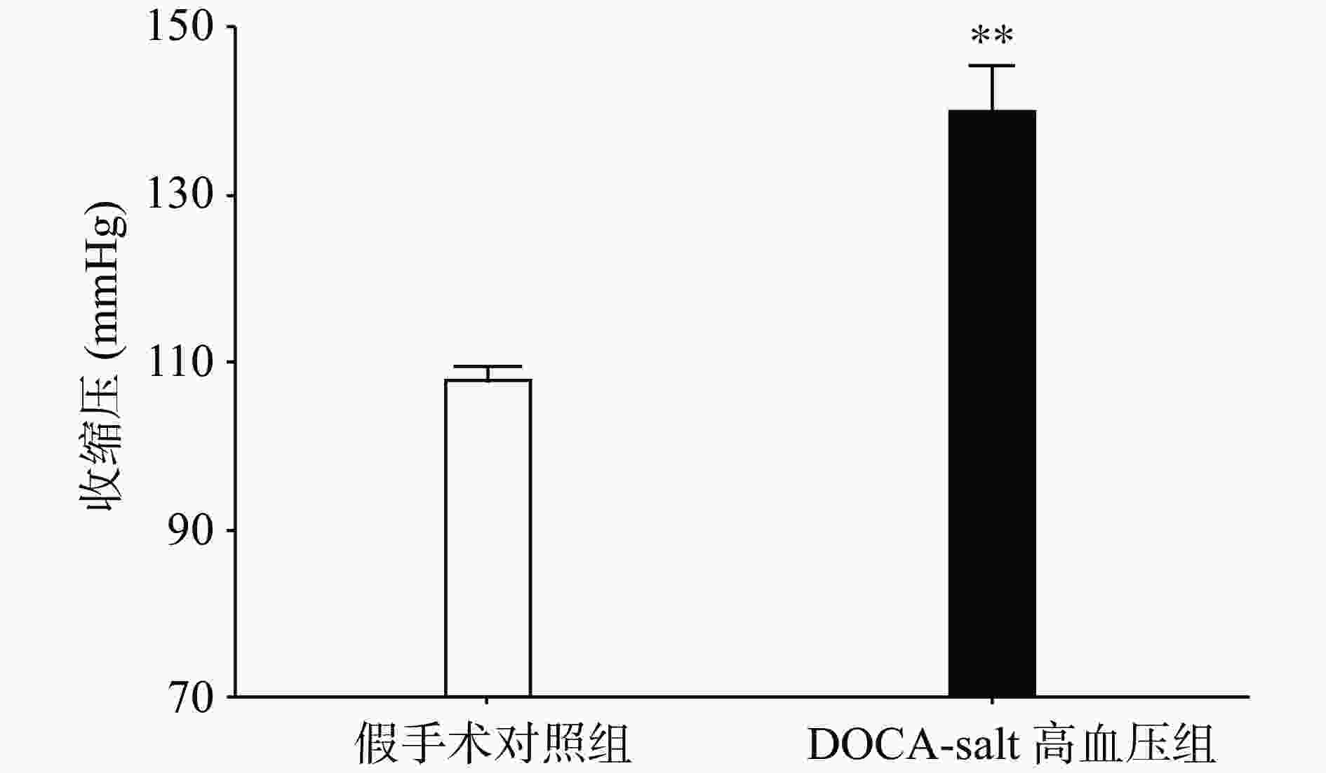
    • 手术后分别测量高血压模型组和假手术组动物的血压水平,发现与假手术组相比,高血压模型组动物的收缩压水平显著上升(P<0.01,图1)。

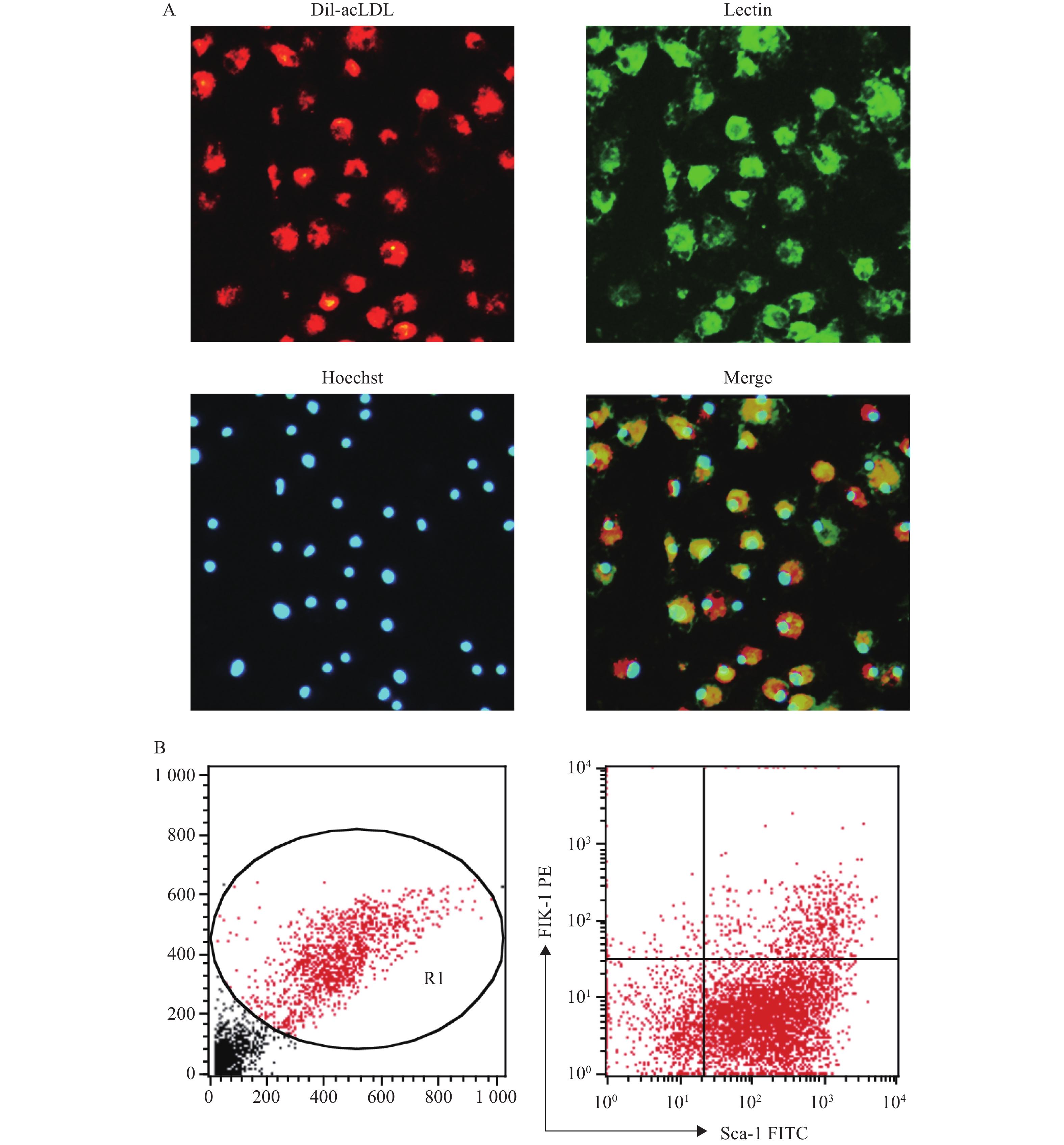
    • 将培养至第7天的骨髓来源的EPC通过荧光显微镜法鉴定,显示Dil-acLDL/Lectin/Hoechst 33258三染阳性的细胞的比例为87.18%;通过流式细胞仪法鉴定,显示Sca-1和Flk-1双标阳性的细胞比例为(11.2±1.5):1,见图2。以上结果与文献报道中使用同类鉴定方法的结果相近[7-10]

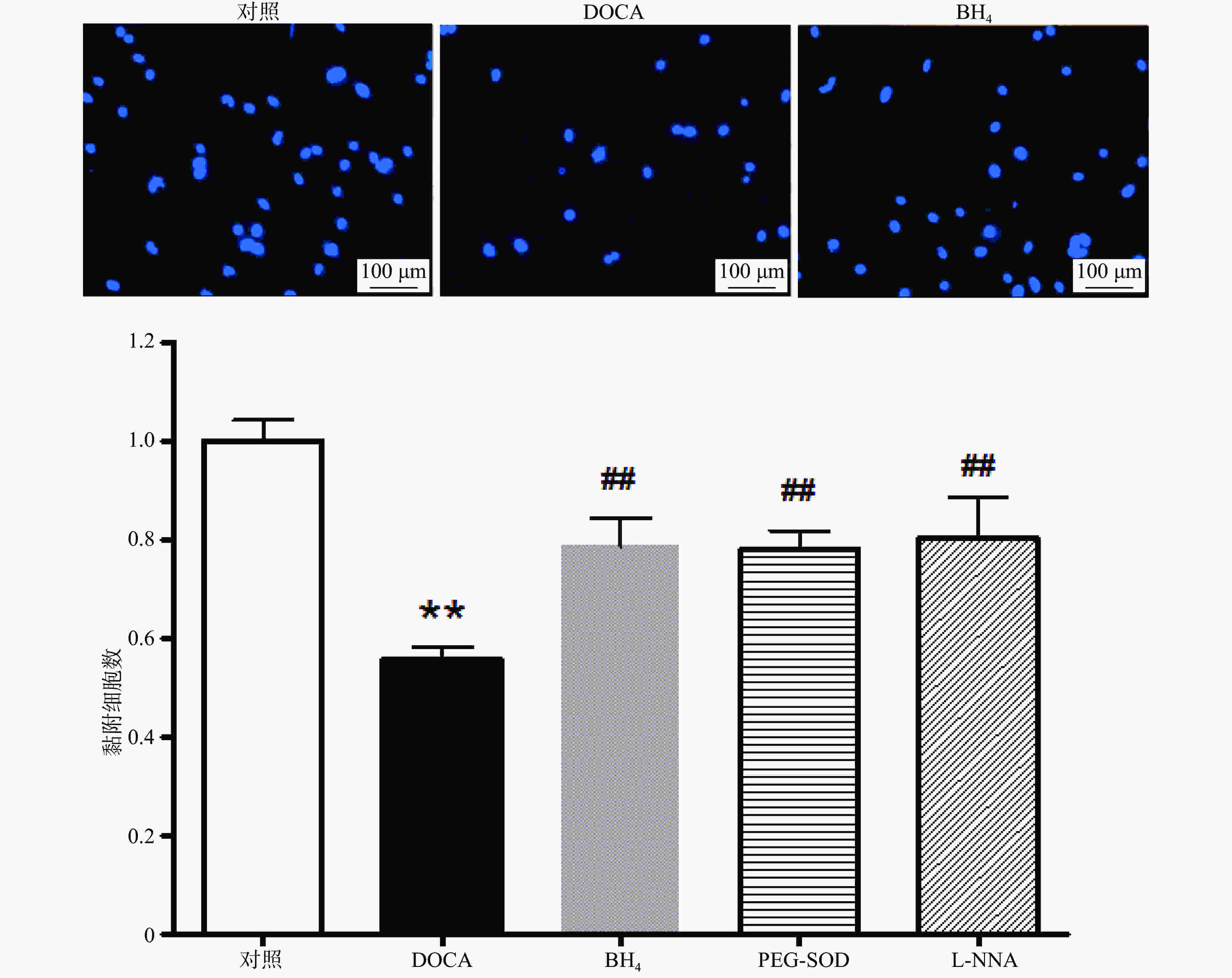
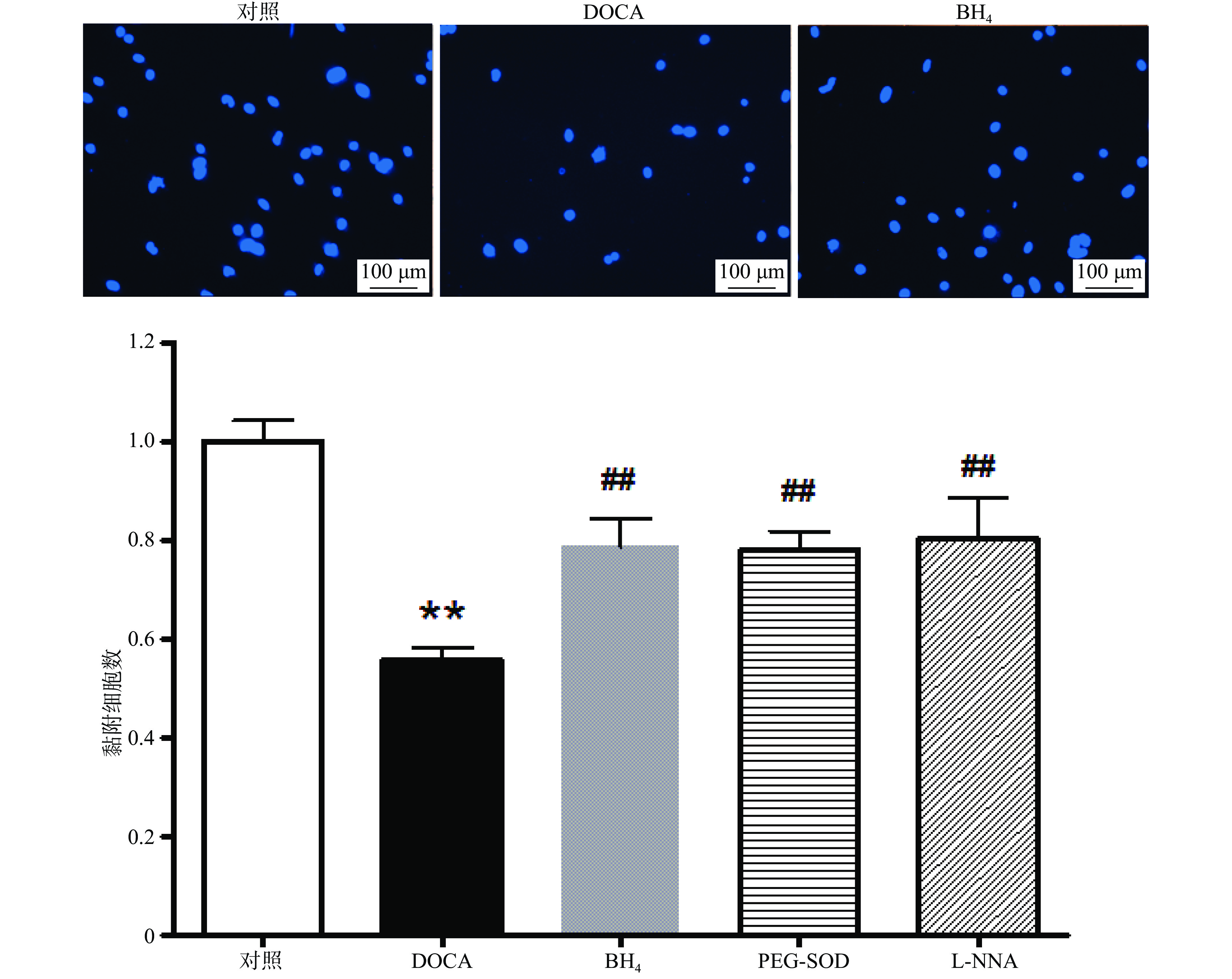
    • 与对照组相比,DOCA-salt高血压小鼠EPC的黏附功能受损,而分别用BH4 (10 µmol/L), PEG-SOD (100 U/L),L-NNA (0.8 mmol/L)3种药物孵育24 h后,其黏附功能得到显著改善(图3)。

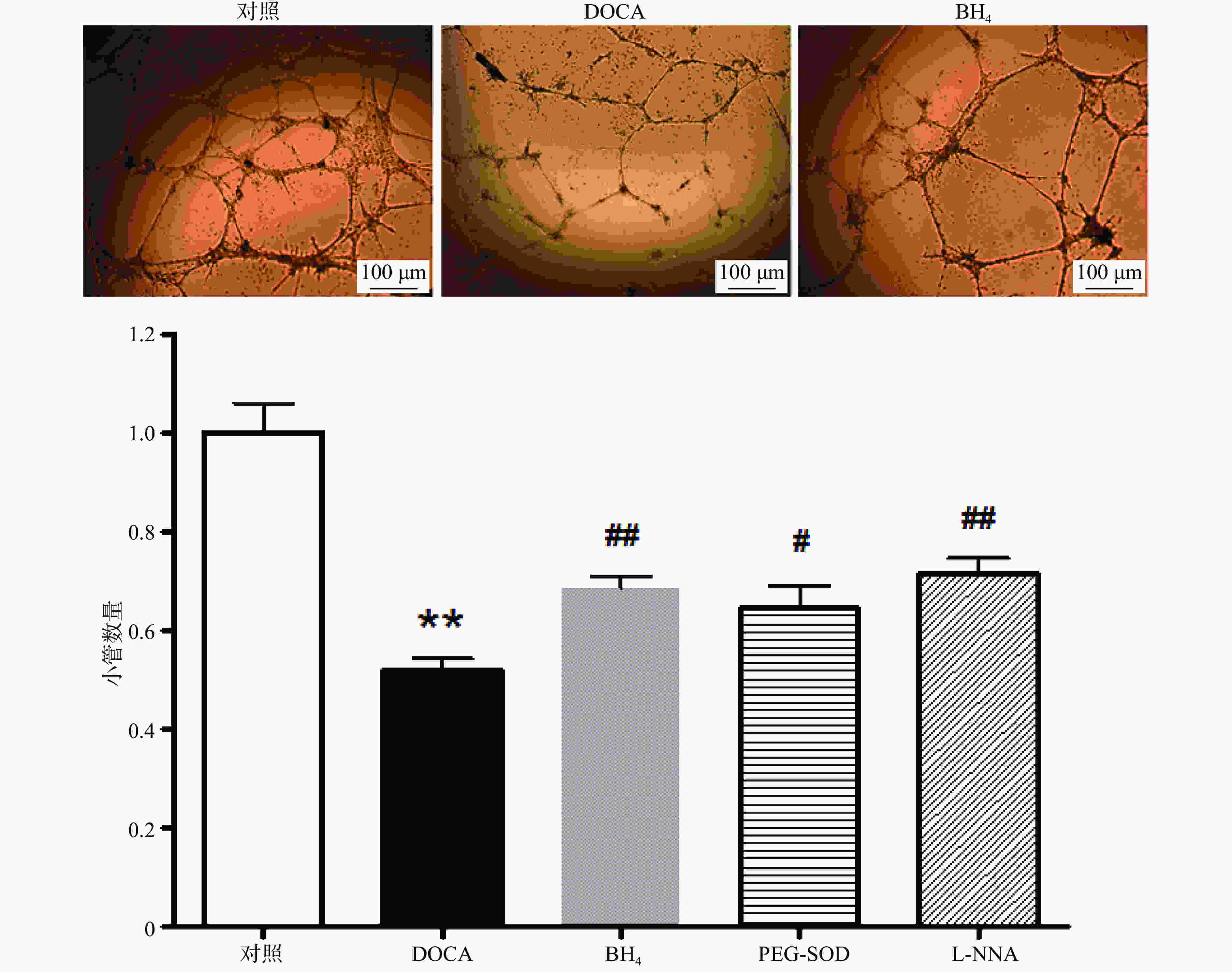
    • 与对照组相比,DOCA-salt高血压小鼠EPC的小管形成功能受损,而分别用BH4 (10 µmol/L), PEG-SOD (100 U/L),L-NNA (0.8 mmol/L)3种药物孵育24 h后,其小管形成功能得到显著改善(图4)。

    • 本研究发现,BH4、PEG-SOD和L-NNA均可有效改善DOCA-salt高血压小鼠EPC受损的功能。但此3种药物改善DOCA-salt小鼠EPC功能的具体机制尚有待进一步探究。

      近年来研究发现,eNOS除了合成一氧化氮(nitric oxide, NO)外,在某些病理情况下,还是超氧阴离子(O2-)的重要来源。BH4是eNOS的必要辅助因子,影响NO和O2-的生成。BH4充足时eNOS催化底物L-精氨酸和O2-生成L-胍氨酸和NO;BH4缺乏时,eNOS则发生脱偶联,主要催化O2-产生[5-6, 11]。BH4缺乏是高血压、糖尿病等疾病中内皮功能异常的重要原因[11]

      本课题组和他人曾利用DOCA-salt高血压动物进行过大量血管生物学研究[4-6, 12],发现该动物血管功能异常与血管组织eNOS脱偶联有关。另外首次发现,DOCA-salt高血压小鼠EPC中BH4水平显著下降,出现eNOS脱偶联,而且eNOS脱偶联所引起的O2-水平升高是导致EPC功能异常的重要机制之一[5]。接着,我们还尝试采用一些药物手段对eNOS脱偶联相关的信号通路/分子进行了干预:①通过外源性补充BH4促进EPC的eNOS复偶联;②用L-NNA抑制脱偶联的eNOS活性,使其产生的O2-水平降低;③补充PEG-SOD直接清除EPC中升高的O2-[5]。我们发现,这些药物干预手段都能显著降低DOCA-salt高血压小鼠EPC的O2-水平[5]。本研究在这些发现的基础上进一步证实,这3种药物干预手段也能显著改善DOCA-salt高血压小鼠EPC的黏附功能和小管形成功能。这些结果也反过来验证了DOCA-salt小鼠EPC功能异常与eNOS脱偶联的关系。

      通过本研究证实,BH4、PEG-SOD、L-NNA均可有效改善DOCA-salt高血压小鼠EPC受损的功能。这些研究结果可为高血压血管并发症的治疗提供参考,也可为改善高血压EPC移植治疗效果提供实验基础。

Reference (12)

Catalog

    /

    DownLoad:  Full-Size Img  PowerPoint
    Return
    Return